
Spinal fusion procedures depend on the precise placement of pedicle screws to stabilize the vertebral column. Because these screws are inserted directly adjacent to the spinal canal and the exiting nerve roots, even a minor misplacement can have serious neurological consequences. Pedicle screw testing is a targeted intraoperative neuromonitoring (IONM) protocol that gives surgical teams immediate, objective feedback on whether every screw placed is safely within bone — and away from the nerves.
What Pedicle Screw Testing Is
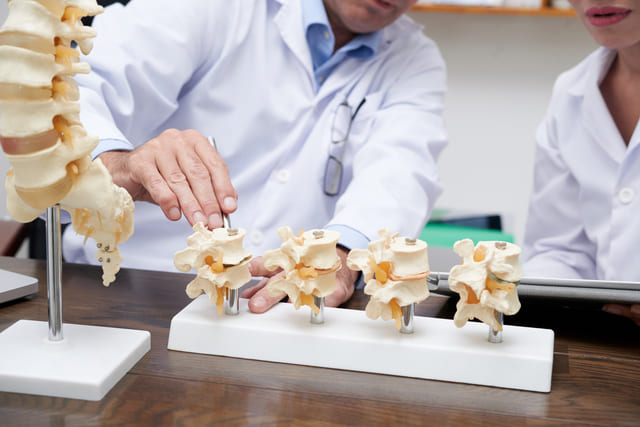
Pedicle screw testing is a specialized application of Triggered EMG. During spinal stabilization, surgeons place titanium screws into the pedicles — the dense bony bridges of the vertebrae that sit directly beside the spinal canal and the nerve roots exiting at each level. Once a screw is seated (or while the pilot hole is being prepared), the neuromonitoring specialist uses a hand-held probe to deliver a precise, controlled electrical current directly to the top of the screw.
The system then measures the electrical threshold — in milliamperes (mA) — required to produce a muscle contraction in the patient's legs or arms.
Why It Matters in Surgery
Detecting Bone Breaches in Real Time Bone is a natural insulator. When a pedicle screw is correctly contained within the bone, a relatively high amount of electrical current is required to stimulate the adjacent nerve — typically above 15 to 20 mA. When a screw has cracked or breached the thin bone wall surrounding the pedicle, that current takes the path of least resistance directly to the nerve. A muscle response triggered at a low threshold — typically below 8 to 10 mA — gives the team an immediate, objective warning that the screw is dangerously close to or in contact with a nerve root.
Correcting Misplacement Before Surgery Is Complete A misplaced pedicle screw can compress or lacerate a nerve root, leading to severe post-operative complications including chronic pain, numbness, radiculopathy, or foot drop. Pedicle screw testing identifies a medial or lateral breach within seconds of hardware placement, while the patient is still on the table and the surgeon can act. The screw can be backed out and redirected immediately, before any permanent injury occurs.
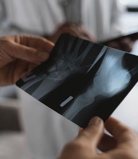
Filling the Gap That Imaging Leaves Intraoperative X-ray and fluoroscopy provide a visual snapshot of where a screw sits anatomically — but they cannot reliably detect a microscopic bone breach or predict whether nearby nerve tissue is being irritated. Pedicle screw testing adds a functional, electrical dimension to hardware verification, confirming not just the physical position of the screw but its neurological safety as well.
Hardware Confidence, Case After Case
Pedicle screw testing is a standard component of Texas IOM's monitoring protocol for spinal fusion procedures. Combined with Free-Run EMG, SSEPs, and MEPs, it forms part of a comprehensive intraoperative monitoring program designed to verify both the neurological status of the patient and the safety of the hardware being placed.
Texas IOM's CNIM-credentialed technologists are experienced across a wide range of spinal procedures and work directly with your surgical team to ensure smooth, efficient monitoring from incision to close.

GET STARTED
Ready to Schedule a Case or Learn More?
Our team is easy to reach. Whether you are scheduling your first case or want to learn more about what we do, we are happy to talk.
© Copyright 2026 | All Rights Reserved Texas IOM | Terms & Conditions | Privacy Policy