
Protecting the spinal cord during surgery is only part of the neurological picture. Many of the most serious intraoperative nerve injuries occur at the peripheral level — in individual nerves far from the cord — or along the cranial nerves that emerge directly from the brain. Texas IOM provides Nerve Conduction Studies (NCS) and Cranial Nerve Monitoring (CNM) as essential components of a complete intraoperative neuromonitoring (IONM) program.
Nerve Conduction Studies (NCS)
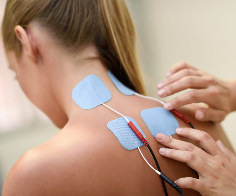
An intraoperative Nerve Conduction Study measures the speed and strength of an electrical impulse traveling through a specific peripheral nerve. Stimulating electrodes are placed at one point on the nerve and recording electrodes are positioned further down the pathway, giving the team an immediate assessment of how efficiently that nerve is transmitting signals.
NCS is particularly valuable during complex orthopedic reconstructions, peripheral nerve surgeries, and trauma cases where nerves are at risk of being stretched, compressed, or entrapped. It provides direct, real-time feedback on whether a nerve's functional integrity is being compromised by a surgical maneuver.
NCS also allows the monitoring team to establish a clear baseline of nerve health at the start of the procedure. This makes it possible to distinguish between pre-existing damage — such as chronic neuropathy — and acute stress introduced during the surgery, ensuring that any new change is identified and addressed immediately.
Cranial Nerve Monitoring (CNM)
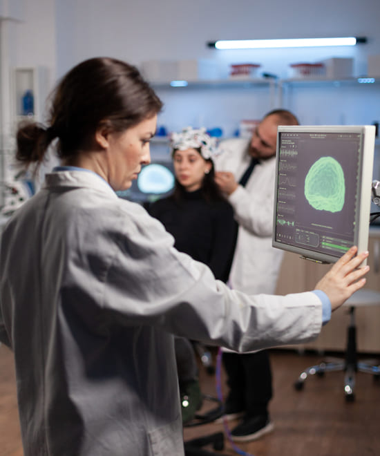
Cranial Nerve Monitoring focuses on the 12 specialized nerves that emerge directly from the brain and brainstem, each controlling a distinct and vital function. This modality typically combines spontaneous electromyography (EMG) — which continuously listens for warning activity when a nerve is disturbed — with triggered stimulation to explicitly verify a nerve's location and pathway.
In practice, electrodes may be placed in the facial muscles to monitor the Facial Nerve (CN VII), in the vocal cords to track the Vagus Nerve (CN X), or in other targeted muscle groups depending on the procedure and the cranial nerves at risk.
During skull base surgeries, brainstem tumor resections, complex ENT procedures, and thyroid surgeries, cranial nerves are frequently microscopic, displaced by tumors, or obscured by scar tissue. CNM acts as a functional map, allowing the surgeon to precisely locate and identify nerve tissue before making an incision. Because cranial nerves control essential functions — including facial movement, eye control, hearing, swallowing, and speech — real-time CNM alerts provide immediate warning if a nerve is being stressed, giving the surgeon the opportunity to adjust before any permanent deficit occurs.
Targeted Monitoring for Complex Procedures
The combination of NCS and Cranial Nerve Monitoring extends the protective reach of IONM beyond the spinal cord and into the peripheral and cranial nerve systems where some of the most consequential intraoperative injuries take place. Texas IOM's team applies these modalities as part of a procedure-specific monitoring plan developed in coordination with your surgical team.

GET STARTED
Ready to Schedule a Case or Learn More?
Our team is easy to reach. Whether you are scheduling your first case or want to learn more about what we do, we are happy to talk.
© Copyright 2026 | All Rights Reserved Texas IOM | Terms & Conditions | Privacy Policy