
While modalities like EEG and evoked potentials focus on large neural pathways and global brain function, protecting individual nerve roots in real time requires a more localized approach. Electromyography (EMG) serves as the most direct sensor available for detecting mechanical nerve irritation the moment it occurs in the operating room.
Texas IOM utilizes EMG in two distinct and complementary forms: Free-Run (Spontaneous) EMG and Triggered EMG.
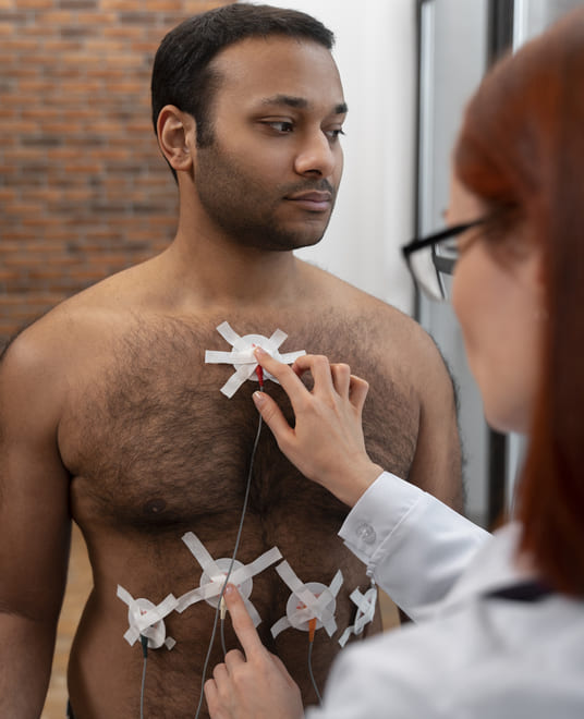
Free-Run (Spontaneous) EMG
Free-Run EMG is a passive, continuous recording of the electrical activity occurring naturally within specific target muscles. Recording electrodes are placed in muscle groups corresponding to the nerve roots at risk during surgery. Without any external stimulation, the system listens constantly to the baseline activity of those nerves throughout the procedure.
Under general anesthesia, healthy, resting muscles are electrically silent. If a surgeon touches, stretches, compresses, or applies traction to a nerve root — even briefly and inadvertently — the irritated nerve fires an immediate burst of electrical impulses to its target muscle. Free-Run EMG captures this activity in real time, generating both a visual waveform on the monitoring screen and an audible alert in the operating room. The moment the surgical team hears that signal, they know a nerve is under mechanical stress and can release traction immediately — before a temporary irritation has any opportunity to become permanent nerve damage.
Triggered EMG
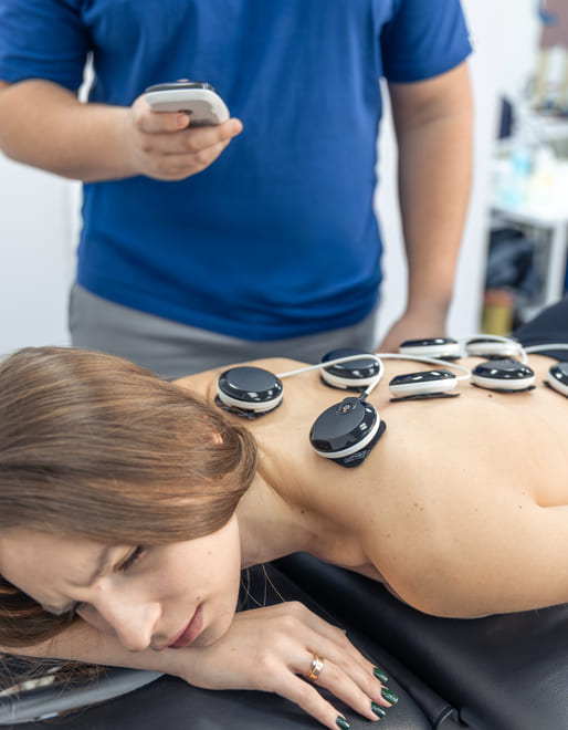
Unlike the passive nature of Free-Run monitoring, Triggered EMG is an active diagnostic test. The neuromonitoring specialist or surgeon uses a hand-held stimulating probe to deliver a precise electrical pulse directly to a specific structure or piece of tissue. The system then measures whether that stimulus produces a corresponding electrical response in a connected muscle group downstream.
This technique has two primary applications in the operating room:
Pedicle Screw Verification: One of the most common uses of Triggered EMG is confirming the safe placement of pedicle screws during spinal fusion. Because bone is a natural electrical insulator and a breached bone wall conducts electricity easily, the surgeon can stimulate the top of a placed screw. A muscle contraction triggered at a low electrical threshold signals that the screw has breached the bone wall and is near or contacting a nerve root — giving the surgeon the chance to redirect it immediately.
Tissue Identification and Nerve Mapping: In surgeries where tumor tissue or scar tissue has engulfed delicate nerve structures, visual identification alone is not reliable. A triggered probe can map the surgical field, identifying exactly where functional nerve tissue is located so that the surgeon can resect tumor without cutting an essential nerve.
A Dual Approach to Peripheral Nerve Safety
Free-Run EMG acts as a continuous, silent sentry that sounds an alarm the moment a nerve is disturbed. Triggered EMG serves as an active navigational tool for testing structures and confirming safe surgical margins. Together, they form a layered defense that significantly reduces the risk of localized muscle weakness, foot drop, or paralysis following complex spinal and peripheral nerve procedures.

GET STARTED
Ready to Schedule a Case or Learn More?
Our team is easy to reach. Whether you are scheduling your first case or want to learn more about what we do, we are happy to talk.
© Copyright 2026 | All Rights Reserved Texas IOM | Terms & Conditions | Privacy Policy